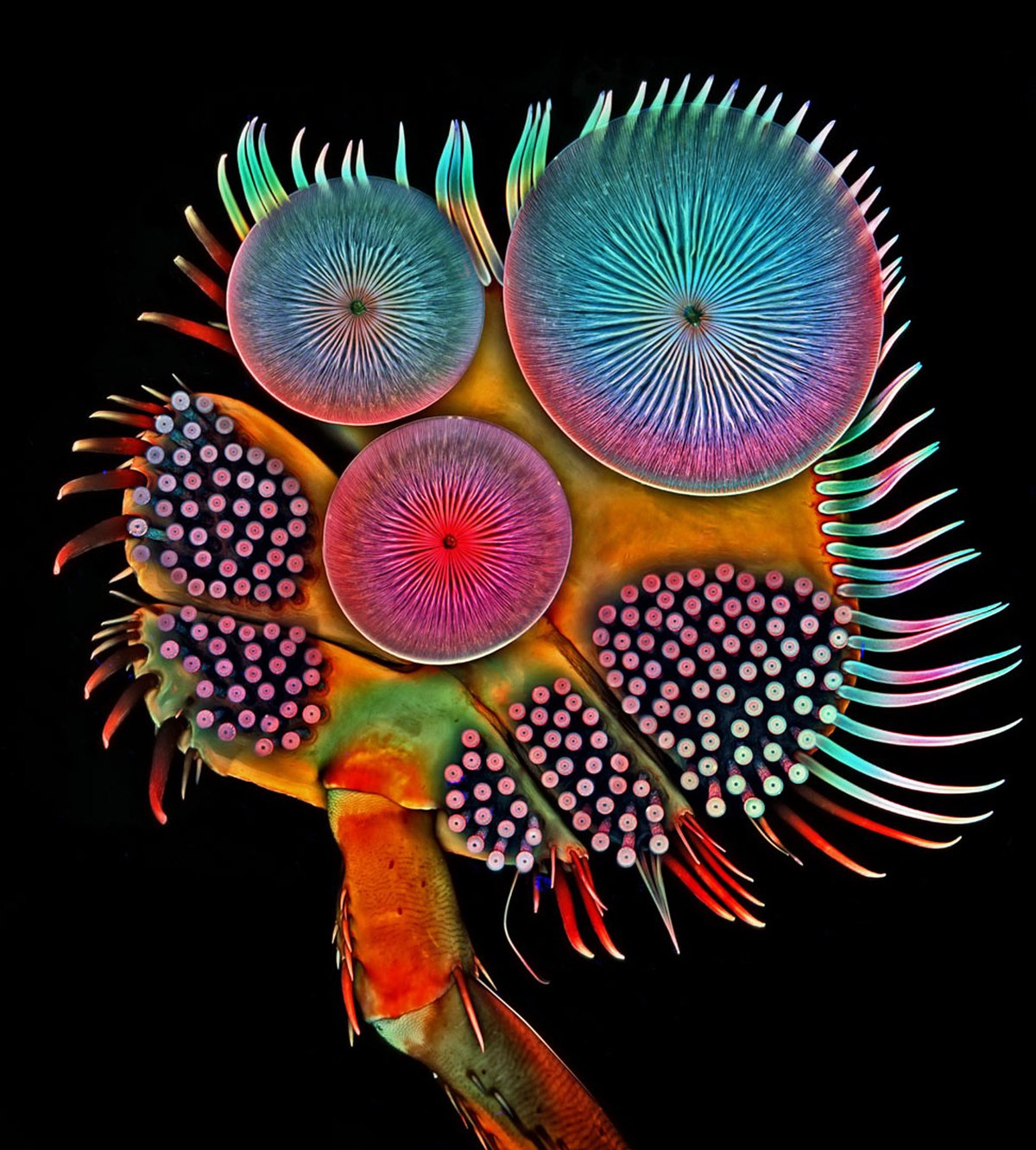
Have you ever wondered what a male diving beetle’s foot looks like under a microscope? Probably not, but it reveals itself to be a fascinating abstract composition of vibrant colours—turquoise, magenta, purple, red—in an image taken by the scientist and photographer Igor Siwanowicz. The work snagged fifth place in the 2016 Nikon Small World Photomicography Competition, launched in 1975 by the Japanese camera manufacturer, which judges photographs of objects magnified by a light microscope. The Bruce Museum in Greenwich, Connecticut is showing the top 20 entries in the 2016 competition (until 29 October), which include subjects such as dyed neurons from human skin cells (Rebecca Nutbrown, third place) and a zebrafish embryo (Oscar Ruiz, first place). The Bruce is one of nine North American venues to show photographs from the competition, but has also added an exhibit of 20th-century microscopes used by Edward Bigelow and Paul Howes, former directors of the museum.


